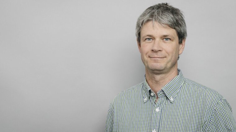
Prof. Rainer Heintzmann.

Biological Nanoimaging

Prof. Dr. Rainer HEINTZMANN
Prof. Rainer Heintzmann.
Image: Sven Döring/Leibniz-IPHT
Rainer HEINTZMANN
Rainer HEINTZMANN
Image: Sven Döring, IPHT Jena.

Prof. Dr. Rainer HEINTZMANN

Email: rainer.heintzmann@uni-jena.de
Phone: +49 3641-9-48350

As a professor of physical chemistry, Rainer Heintzmann is a member of the Faculty of Chemistry and Earth Sciences and the Faculty of Physics and Astronomy. He also heads a research unit at the Leibniz Institute of Photonic Technology in Jena. His main research direction has been high resolution optical microscopy and microscopy image processing.

The group develops techniques to measure multidimensional information in small biological objects such as cells, cellular organelles, molecules or other structures of interest. We seek to unravel how molecules interact in living cells at specific places (e.g. inside organelles) and at well-defined times (e.g. after stimulation with other molecules). To reach this goal, we make use of molecules that can be switched between different fluorescent states by illumination at separate wavelengths. The transitions between the states can be driven into saturation and the arising non-linear dependencies can be used, e.g. by a technique called structured illumination, to reach a theoretically unlimited optical resolution.

Research Areas

The research of his group concentrates on the improvement of optical microscopy. Specific focus is currently placed on: 

  • Modern optical microscopy for biomedical research
  • Research and implementation of super-resolved fluorescence microscopy through linear and non-linear structured illumination (SIM) and localization microscopy (dSTORM, DNA-PAINT)
  • Hyper-spectral Raman-imaging for combined spatial and spectral information in medical research
  • Affordable microscopes based on 3D-printig and smartphones for education and research
  • Advanced image processing in microscopy
  • Solving inverse problems in microscopy (e.g. image deconvolution and 3D-reconstruction)

Teaching Fields

Prof. Heintzmann teaches undergraduate courses in physical chemistry, as well as specialized courses on optics and image processing. These currently include:

  • Light microscopy
  • Biophotonics
  • Image processing in microscopy

Research Methods

The methods developed in Prof. Heintzmann’s laboratories are applied, in collaboration with biologists, to biomedical problems. Available methods are:

  • Fast structured illumination microscopy
  • Localization microscopy (dSTORM, DNA-PAINT)
  • Confocal microscopy
  • Hyper-spectral Raman microscopy
  • Affordable and flexible microscopes through 3D-printing and off-the-shelf electronics

Recent Research Results

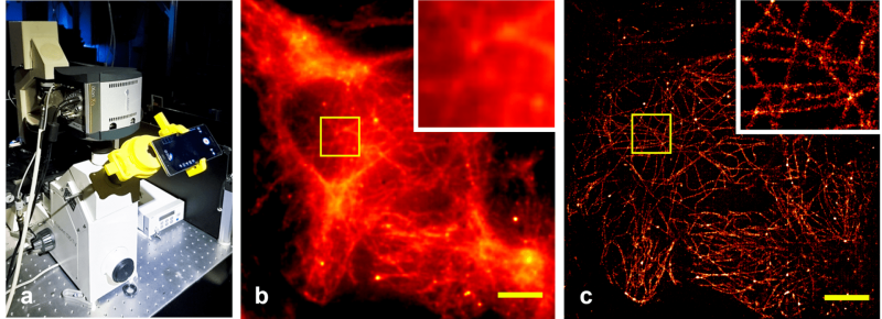
CellSTORM - affordable localisation microscopy on a smartphone.
Image: Heintzmann research group

The Nanobiophotonics group has a long tradition working on aspects of structured illumination. It remains a strong focus of research in our group. Recently, in cooperation with the group of Dr. Thomas Ach, we have used our SIM-system for fast, multi-color imaging of agerelated macula-degeneration (AMD) [1]. Optimization of SIMreconstruction remains an important topic for us [2,3], e.g., related to identification and avoidance of motion artifacts [4]. Another line of research concentrates on pointillistic or single-molecule localization (SML)-based imaging modes. With dSTORM and DNA-PAINT, we use two SML methods, which rely on the precise localization of individual fluorescent dyes in the sample and are able to resolve structural details below 25 nm. In cooperation with external partners, these methods are employed e.g. in cancer research. We currently also develop approaches for a further resolution improvement through novel holographic illumination techniques [5], as  well as sequential staining techniques for super-resolved imaging of many different colors. Spectroscopy based on Raman-scattering offers high molecular specificity without the necessity for staining or other marking steps. However, the integration into normal confocal imaging modalities limits speed and quality of the measurement due to low efficiency and heating effects [6]. We are thus working on hyperspectral Raman-imaging by combining integral-field spectrometry with light-sheet illumination. Our system enables parallel recording of 50×50 spectra, reaching a 50-fold enhancement in speed compared to standard confocal systems [7]. A similar method is employed in the visible spectral range for studies on AMD. Our project UC2 [8] is an open-source general-purpose modular framework for making interactive projects with optics and electronics. Relying on the basic principles makes UC2 particularly easy to use and flexible to adapt for a wide range of applications. Building an optical setup is simplified using 3D-printed cubes, each holding a specific component (e.g. lens, mirror) on a magnetic square-grid baseplate. The use of widely available consumables, together with open documentation and software, offers an extremely low-cost and accessible alternative for both education and research areas. The UC2 concept serves as a base for building microscopes and thanks to 3D printing it is ideal for rapid. The examples of many of the applications and some results are shown in the UC2 manuscript [9].

[1] Mohammed et al., Vision 2, 38 (2018).
[2] Karras et al., Opt. Commun. 69, 436 (2019).
[3] Ingerman et al., Journal of Microscopy 3, 273 (2019).
[4] Förster et al., Opt. Express 26, 20680 (2018).
[5] Jügler et al., OSA Technical digest paper FM4E.5 (2018).
[6] Hauswald et al., PLoS ONE 14, e0220824 (2019).
[7] Zegarra-Valverde et al., OSA Technical Digest, paper FM3F.5d (2019).
[8] UC2 GitHub repository [Online], https://github.com/bionanoimaging/UC2-GIT (2020).
[9] Diederich et al., Nature Commun. 11, 5979 (2020).
[10] Diederich et al., PloS One 14, e0227096 (2019).
[11] Diederich et al., bioRxiv 2020.09.04.283085.

link to the Biological Nanoimaging GroupExternal link